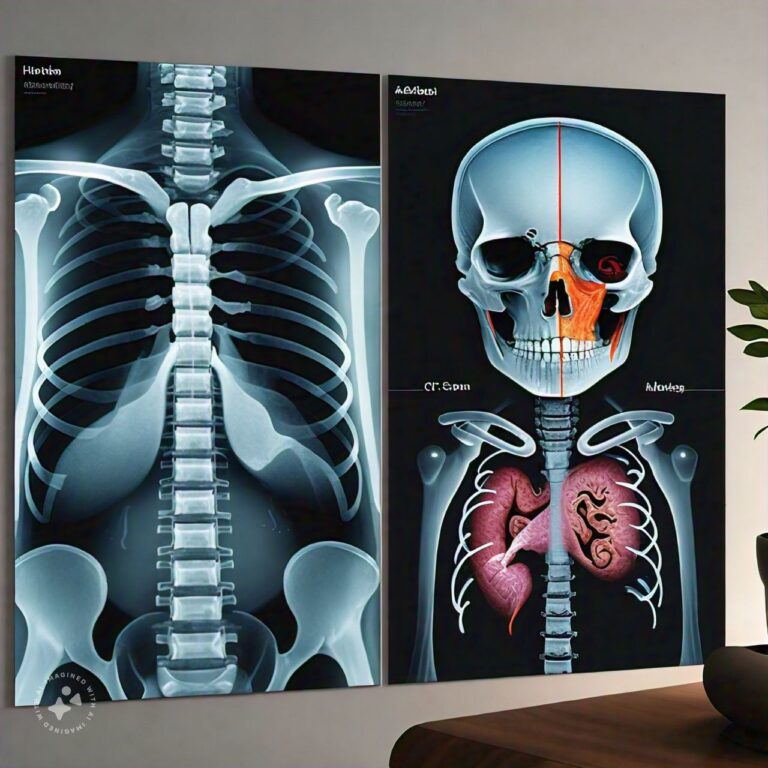
Differences Between X-ray and CT Scan
X-rays and CT scans are both diagnostic imaging tools that help doctors view the internal structures of the body. While they serve a similar purpose, they differ in how they work and the level of detail they provide. X-rays are a more basic and widely used imaging technique that provides two-dimensional images, mainly useful for examining bones, teeth, and the chest. CT scans (Computed Tomography), on the other hand, use multiple X-rays taken from different angles and computer processing to create more detailed cross-sectional images, allowing for a better look at soft tissues, blood vessels, and complex areas of the body.
Choosing between an X-ray and a CT scan depends on what part of the body needs to be examined and the level of detail required for diagnosis. Understanding the key differences between X-rays and CT scans, their uses, and their advantages can help patients and healthcare providers make informed decisions about diagnostic imaging.
X-ray Overview
Introduction to X-rays
An X-ray is a type of electromagnetic radiation that passes through the body to create images of the internal structures, primarily focusing on bones and dense tissues. X-rays are among the most common and oldest forms of medical imaging. They were first discovered by Wilhelm Conrad Roentgen in 1895 and have since become an essential tool in diagnosing a variety of medical conditions.
X-rays work by directing a controlled beam of radiation through the body, with different tissues absorbing the X-rays to varying degrees. Dense materials like bones absorb more radiation and appear white on the resulting image, while less dense materials, like soft tissues, allow more X-rays to pass through and appear darker on the image.
How X-rays Work
X-ray imaging works on the principle that different tissues in the body absorb X-rays to varying degrees, depending on their density. When an X-ray machine sends a controlled amount of radiation through the body, the rays pass through soft tissues (like muscles and organs) but are absorbed by denser structures (like bones and teeth). This difference in absorption creates a contrast on the X-ray film or digital detector, which results in a two-dimensional image.
- Bones and Teeth:
Bones and teeth are dense and absorb the majority of the X-rays, making them appear white or light gray on the image. - Soft Tissues and Organs:
Soft tissues, such as muscles, fat, and internal organs, absorb fewer X-rays and appear in shades of gray. - Air:
Air-filled structures, like the lungs, allow X-rays to pass through easily, making them appear darker or black on the image.
Common Uses of X-rays
X-rays are used in a wide range of medical diagnoses, particularly for examining bones, teeth, and certain organs. Common uses include:
- Bone Fractures:
X-rays are frequently used to detect fractures or broken bones. They provide clear images of the bone structure, making it easy to identify cracks, breaks, or dislocations. - Dental Imaging:
Dentists use X-rays to check for cavities, tooth decay, and issues with tooth alignment or jawbone structure. - Chest X-rays:
Chest X-rays are used to examine the lungs, heart, and chest cavity. They can help diagnose conditions such as pneumonia, tuberculosis, lung cancer, and heart enlargement. - Mammography:
X-rays are used in mammography to detect breast cancer by providing images of breast tissue. - Infection and Arthritis:
X-rays can reveal signs of infection, inflammation, or degenerative conditions like arthritis, where joint spaces may become narrowed or show bone erosion.
Benefits of X-rays
X-rays offer several advantages, particularly in terms of speed, accessibility, and the ability to diagnose common conditions. Some of the key benefits include:
- Quick and Non-invasive:
X-rays are fast and painless. The process usually takes just a few minutes, and the patient experiences minimal discomfort during the procedure. - Widely Available:
X-rays are available in most healthcare facilities, including hospitals, urgent care centers, and dental offices, making them accessible to most patients. - Low Cost:
Compared to more advanced imaging techniques like CT or MRI, X-rays are relatively inexpensive, making them a cost-effective option for basic diagnostic imaging. - Good for Bone Imaging:
X-rays are especially useful for examining the bones and joints, providing detailed images of fractures, dislocations, and other skeletal issues.
Risks and Limitations of X-rays
Although X-rays are a valuable diagnostic tool, they do come with certain risks and limitations:
- Radiation Exposure:
X-rays involve exposure to ionizing radiation, which can increase the risk of cancer with repeated exposure. However, the level of radiation in most X-rays is low, and the benefits of obtaining diagnostic information usually outweigh the risks. - Limited Soft Tissue Detail:
X-rays are not ideal for imaging soft tissues like the brain, muscles, or internal organs, as these tissues do not absorb X-rays well and appear faint on the images. - Two-Dimensional Images:
X-rays provide flat, two-dimensional images, which may not offer enough detail for complex diagnoses, particularly when examining overlapping structures. - Pregnancy Considerations:
X-rays are generally avoided during pregnancy due to the risk of radiation exposure to the developing fetus, although exceptions may be made in emergency situations.
CT Scan Overview
Introduction to CT Scans
CT scans (Computed Tomography), also known as CAT scans, are a more advanced form of X-ray imaging that creates detailed, cross-sectional images of the body. A CT scan uses multiple X-ray beams from different angles and combines them with computer processing to generate 3D images of internal structures. This allows for a much more detailed examination of bones, soft tissues, blood vessels, and organs than a standard X-ray.
CT scans are often used when more detailed information is needed to diagnose complex conditions, especially when the condition involves soft tissues, such as the brain, lungs, or abdomen. CT scans are also invaluable in emergency situations for detecting internal injuries, bleeding, or other life-threatening conditions.
How CT Scans Work
CT scans work by taking multiple X-ray images of a particular part of the body from different angles. During a CT scan, the patient lies on a table that slides through a large, doughnut-shaped machine called a gantry. The gantry contains an X-ray tube that rotates around the patient, taking multiple X-ray images as it moves.
These images are then processed by a computer, which reconstructs them into detailed cross-sectional "slices" of the body. These slices can be viewed individually or stacked together to create a 3D image of the scanned area.
The resulting images provide far more detail than a standard X-ray, making CT scans ideal for detecting internal injuries, tumors, infections, and other complex conditions.
Common Uses of CT Scans
CT scans are often used when more detailed information is needed than what an X-ray can provide. Some common uses of CT scans include:
- Detecting Internal Injuries:
CT scans are particularly valuable in emergency situations, such as after trauma or accidents, to detect internal bleeding, organ damage, or fractures that may not be visible on a standard X-ray. - Diagnosing Tumors and Cancers:
CT scans can provide detailed images of tumors, making them useful in diagnosing cancers in the brain, lungs, abdomen, and other parts of the body. - Evaluating Brain and Spinal Conditions:
CT scans of the brain can help diagnose conditions such as stroke, brain tumors, aneurysms, and traumatic brain injuries. They can also assess spinal injuries and disorders. - Examining Blood Vessels (CT Angiography):
CT scans can be used to visualize blood vessels and detect blockages, aneurysms, or other vascular abnormalities. This is often done using a contrast agent that highlights the blood vessels. - Abdominal and Pelvic Conditions:
CT scans are commonly used to diagnose conditions affecting the abdominal organs, such as appendicitis, kidney stones, liver disease, and bowel obstructions.
Benefits of CT Scans
CT scans offer several advantages over standard X-rays, particularly when it comes to providing detailed and comprehensive images of the body's internal structures. Key benefits include:
- Detailed Cross-sectional Images:
CT scans provide detailed cross-sectional images of the body, allowing for better visualization of complex structures, including bones, soft tissues, and blood vessels. - 3D Reconstruction:
CT scans can produce three-dimensional images of the body, providing a more complete view of the area being examined. This is particularly useful for surgical planning or assessing complex fractures. - Comprehensive Diagnostic Tool:
CT scans are a valuable tool for diagnosing a wide range of conditions, including cancer, trauma, infections, and vascular diseases. - Quick and Non-invasive:
Like X-rays, CT scans are non-invasive and can be performed quickly, often in just a few minutes.
Risks and Limitations of CT Scans
While CT scans provide detailed diagnostic information, they also come with some risks and limitations:
- Higher Radiation Exposure:
CT scans involve higher doses of radiation compared to standard X-rays. Repeated CT scans over time can increase the risk of cancer, especially in younger patients or those who require frequent imaging. - Use of Contrast Agents:
Some CT scans require the use of a contrast agent (usually iodine-based) to enhance the visibility of certain tissues or blood vessels. In rare cases, patients may have allergic reactions to the contrast agent, and it can be harmful to those with kidney problems. - Not Suitable for Pregnant Women:
Due to the higher radiation levels, CT scans are generally avoided in pregnant women unless absolutely necessary to avoid potential harm to the developing fetus. - Cost:
CT scans are more expensive than standard X-rays due to the advanced technology and detailed images they provide.
Differences Between X-ray and CT Scan
- Technology:
- X-ray: Uses a single beam of X-rays to create two-dimensional images of bones and tissues.
- CT Scan: Uses multiple X-ray beams from different angles, combined with computer processing, to create cross-sectional and 3D images of the body.
- Level of Detail:
- X-ray: Provides basic images of bones and some tissues, primarily used for detecting fractures or lung conditions.
- CT Scan: Offers detailed images of bones, soft tissues, blood vessels, and organs, making it ideal for diagnosing complex conditions.
- Radiation Exposure:
- X-ray: Involves lower levels of radiation exposure.
- CT Scan: Involves higher radiation exposure due to the multiple X-ray beams used.
- Use of Contrast Agents:
- X-ray: Typically does not require contrast agents, except in specific cases like barium X-rays for digestive tract imaging.
- CT Scan: Often uses contrast agents to enhance visibility of blood vessels and soft tissues.
- Imaging Scope:
- X-ray: Best for imaging bones, teeth, and the chest. Limited in its ability to show soft tissue details.
- CT Scan: Provides detailed imaging of both bones and soft tissues, such as muscles, organs, and blood vessels.
- Speed:
- X-ray: Quick and typically takes just a few minutes.
- CT Scan: Takes longer than an X-ray but can still be completed in a matter of minutes.
- Cost:
- X-ray: Less expensive, making it a cost-effective option for routine imaging.
- CT Scan: More expensive due to the advanced technology and detailed images.
- Common Uses:
- X-ray: Primarily used for diagnosing fractures, dental issues, and chest conditions like pneumonia.
- CT Scan: Used for diagnosing internal injuries, cancers, soft tissue conditions, and complex bone fractures.
- Suitability for Emergencies:
- X-ray: Useful for quickly diagnosing bone fractures in emergency settings.
- CT Scan: Preferred in emergencies for detecting internal injuries, bleeding, and other life-threatening conditions.
- 3D Imaging:
- X-ray: Provides two-dimensional images.
- CT Scan: Produces detailed 3D images, offering more comprehensive diagnostic information.
Conclusion
In conclusion, X-rays and CT scans are both essential diagnostic tools used in modern medicine, but they differ significantly in their technology, level of detail, and appropriate use cases. X-rays are simple, fast, and cost-effective imaging techniques primarily used to examine bones, teeth, and chest conditions. They provide two-dimensional images and involve lower levels of radiation exposure, making them ideal for routine diagnostics.
CT scans, on the other hand, provide far more detailed and comprehensive images by combining multiple X-rays with computer processing. They are particularly valuable for diagnosing complex conditions involving soft tissues, organs, blood vessels, and internal injuries. However, CT scans come with higher radiation exposure and cost, which makes them less suitable for routine use unless more detailed imaging is required.
Understanding the differences between these imaging techniques allows healthcare providers to choose the most appropriate method based on the patient’s condition, ensuring accurate diagnoses and effective treatment planning.
FAQs
Related Topics
- All
- Animals
- Diseases
- Health
- Money
- Politics
© 2024 OnYelp.com. All rights reserved. Terms and Conditions | Contact Us | About us


