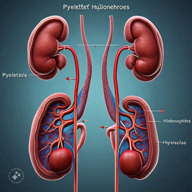
Differences Between Pyelectasis and Hydronephrosis
Pyelectasis and hydronephrosis are two medical conditions that involve the kidneys and urinary system, specifically affecting the renal pelvis, where urine collects before flowing to the bladder. Both conditions refer to the dilation or swelling of the renal pelvis and/or calyces, but they differ in terms of severity, underlying causes, and potential health consequences. Pyelectasis is generally considered a mild or moderate dilation, often observed in prenatal ultrasounds or as a benign finding in adults. Hydronephrosis, on the other hand, represents a more severe dilation, often indicating an obstruction or significant impairment in urine flow. Understanding the distinction between these conditions is crucial for diagnosis, management, and treatment.
Pyelectasis Overview
Pyelectasis refers to the dilation (swelling) of the renal pelvis, the funnel-like structure in the kidney where urine collects before it is funneled into the ureter and eventually the bladder. Pyelectasis is usually detected during prenatal ultrasounds, where it is commonly observed in fetuses, but it can also be found in adults through imaging studies. The term "pyelectasis" is used when the dilation is mild and not necessarily indicative of a serious underlying condition.
Pyelectasis can be either unilateral (affecting one kidney) or bilateral (affecting both kidneys). In many cases, pyelectasis is considered a benign condition, especially when detected prenatally, and may resolve on its own without treatment. However, in some cases, it can be a sign of urinary tract issues, such as obstruction or reflux, that may require monitoring or intervention.
Causes of Pyelectasis:
Pyelectasis can result from a variety of causes, some of which are more serious than others. Common causes include:
- Physiological Pyelectasis in Fetuses:
In many prenatal cases, mild pyelectasis is simply a transient condition caused by the developing urinary system and resolves after birth without any lasting effects. This type of pyelectasis is often considered a variation of normal fetal development. - Urinary Tract Obstruction:
A blockage in the urinary tract, such as a narrowing of the ureter or urethra, can lead to urine buildup in the renal pelvis, causing it to dilate. In such cases, pyelectasis may require closer monitoring to ensure that the obstruction does not lead to more severe complications. - Vesicoureteral Reflux (VUR):
VUR is a condition in which urine flows backward from the bladder into the ureters and sometimes into the kidneys. This can cause the renal pelvis to dilate, leading to pyelectasis. VUR is often diagnosed in children and may resolve with time or require medical management. - Kidney Stones:
The presence of kidney stones can cause partial blockages in the urinary tract, leading to mild dilation of the renal pelvis. If the stone passes on its own, the pyelectasis may resolve without intervention. - Congenital Anomalies:
Some infants are born with structural abnormalities in the urinary tract that can cause pyelectasis. These anomalies may be identified during prenatal ultrasounds or later in life and can sometimes require surgical correction.
Symptoms of Pyelectasis:
In many cases, pyelectasis does not cause noticeable symptoms, particularly when the dilation is mild. When symptoms do occur, they may include:
- Discomfort or Pain:
Some individuals may experience mild discomfort in the abdomen or flank area, particularly if the pyelectasis is caused by an obstruction. - Frequent Urination:
Urinary tract obstruction or VUR can cause increased frequency of urination or urgency. - Recurrent Urinary Tract Infections (UTIs):
Pyelectasis associated with urinary tract abnormalities, such as VUR, can increase the risk of UTIs.
Diagnosis of Pyelectasis:
Pyelectasis is most commonly diagnosed during prenatal ultrasounds, where mild dilation of the renal pelvis may be observed in the developing fetus. In adults or children, imaging studies such as ultrasound, CT scans, or MRI can detect pyelectasis. When pyelectasis is detected, doctors may monitor the condition over time to see if it resolves on its own or if further investigation is needed to determine the underlying cause.
Treatment of Pyelectasis:
The treatment of pyelectasis depends on the underlying cause and the severity of the dilation. In many cases, particularly in prenatal pyelectasis, no treatment is required, and the condition resolves spontaneously. However, if pyelectasis is caused by an obstruction, VUR, or another medical condition, treatment may involve:
- Monitoring:
Mild cases of pyelectasis may simply be monitored with follow-up ultrasounds to ensure that the condition does not worsen. - Surgical Intervention:
If a urinary tract obstruction is causing pyelectasis, surgery may be required to remove the obstruction or correct any structural abnormalities. - Treatment for Reflux or Infection:
In cases of VUR or recurrent UTIs, antibiotics or other medical therapies may be prescribed to manage the condition and prevent complications.
Hydronephrosis Overview
Hydronephrosis is a more severe condition involving the swelling of one or both kidneys due to the buildup of urine, often resulting from an obstruction in the urinary tract. This buildup causes the renal pelvis and calyces (the chambers where urine collects before it moves to the bladder) to become distended, potentially leading to kidney damage if not treated.
Hydronephrosis can affect one kidney (unilateral) or both kidneys (bilateral) and is typically categorized into mild, moderate, or severe based on the degree of swelling. Unlike pyelectasis, which is usually mild and may resolve on its own, hydronephrosis often requires medical intervention to address the underlying cause and prevent long-term damage to kidney function.
Causes of Hydronephrosis:
Hydronephrosis can result from various underlying conditions that block or impede the normal flow of urine. Some of the common causes include:
- Kidney Stones:
Kidney stones can obstruct the flow of urine through the ureters, leading to a backup of urine in the renal pelvis and causing hydronephrosis. - Urinary Tract Obstruction:
Blockages anywhere along the urinary tract, including the ureteropelvic junction (where the kidney connects to the ureter), can lead to the buildup of urine and swelling of the kidney. - Benign Prostatic Hyperplasia (BPH):
In men, an enlarged prostate can obstruct the urethra, making it difficult for urine to flow out of the bladder. This can cause urine to back up into the kidneys, leading to hydronephrosis. - Pregnancy:
During pregnancy, the growing uterus can sometimes press on the ureters, leading to temporary hydronephrosis. This condition often resolves after childbirth. - Vesicoureteral Reflux (VUR):
VUR can cause urine to flow backward from the bladder into the kidneys, leading to hydronephrosis if the reflux is severe. - Tumors or Masses:
Tumors or other masses in the abdomen can compress or obstruct the urinary tract, leading to hydronephrosis.
Symptoms of Hydronephrosis:
The symptoms of hydronephrosis can vary depending on the severity of the condition and whether it is acute or chronic. Common symptoms include:
- Pain in the Flank or Abdomen:
Hydronephrosis often causes pain or discomfort in the side or back, near the affected kidney. - Difficulty Urinating:
An obstruction in the urinary tract can cause difficulty starting urination, reduced urine output, or a sense of incomplete bladder emptying. - Blood in the Urine (Hematuria):
Hydronephrosis caused by kidney stones or other conditions may lead to blood in the urine. - Recurrent UTIs:
Frequent urinary tract infections can be a symptom of hydronephrosis, especially if there is an underlying obstruction or reflux. - Nausea and Vomiting:
In severe cases, hydronephrosis can cause nausea, vomiting, and other symptoms of systemic illness.
Diagnosis of Hydronephrosis:
Hydronephrosis is typically diagnosed using imaging studies, such as ultrasound, CT scans, or MRI. These tests help visualize the kidneys, identify the degree of swelling, and locate any obstructions in the urinary tract. Additional diagnostic tests may include:
- Voiding Cystourethrogram (VCUG):
This test is used to diagnose vesicoureteral reflux by examining how urine flows through the bladder and ureters during urination. - Blood Tests:
Blood tests may be performed to assess kidney function and check for signs of infection or other complications.
Treatment of Hydronephrosis:
The treatment of hydronephrosis depends on the underlying cause and the severity of the condition. Common treatment options include:
- Addressing the Obstruction:
If hydronephrosis is caused by a blockage (such as kidney stones or an enlarged prostate), treatment may involve removing the obstruction through surgery or other medical procedures. - Drainage of the Kidney:
In severe cases of hydronephrosis, a catheter or stent may be placed to drain urine from the kidney and relieve pressure while the underlying cause is treated. - Antibiotics for Infection:
If hydronephrosis is associated with recurrent UTIs, antibiotics may be prescribed to treat or prevent infections. - Surgical Intervention:
Surgery may be required to correct structural abnormalities in the urinary tract or to remove tumors that are causing obstruction.
Differences Between Pyelectasis and Hydronephrosis
- Severity:
Pyelectasis is generally a mild dilation of the renal pelvis and often does not require treatment, whereas hydronephrosis involves more severe swelling and can lead to kidney damage if not addressed. - Underlying Cause:
Pyelectasis is often a transient or benign finding, especially in prenatal cases, while hydronephrosis is typically caused by an obstruction or more serious condition that impairs urine flow. - Symptoms:
Pyelectasis may not cause any noticeable symptoms and is often detected incidentally during imaging studies. Hydronephrosis, on the other hand, can cause significant symptoms, such as pain, difficulty urinating, and recurrent UTIs. - Treatment:
Pyelectasis often requires only monitoring, while hydronephrosis may require medical or surgical intervention to prevent complications and restore normal urine flow. - Long-Term Impact:
Pyelectasis usually resolves on its own without long-term effects, while untreated hydronephrosis can lead to permanent kidney damage and impaired kidney function.
Conclusion
Pyelectasis and hydronephrosis are two related conditions involving the dilation of the renal pelvis, but they differ in terms of severity, underlying causes, and treatment requirements. Pyelectasis is typically a mild, often transient condition that may not cause symptoms or require treatment, especially when detected during prenatal ultrasounds. In contrast, hydronephrosis is a more serious condition that indicates an obstruction or other impairment of urine flow, requiring prompt medical attention to prevent kidney damage. Understanding the differences between these conditions is essential for accurate diagnosis, monitoring, and treatment, ensuring the best outcomes for patients.
FAQs
Related Topics
- All
- Animals
- Diseases
- Health
- Money
- Politics
© 2024 OnYelp.com. All rights reserved. Terms and Conditions | Contact Us | About us


