Differences Between CT Scan and MRI Scan
CT scans (Computed Tomography) and MRI scans (Magnetic Resonance Imaging) are two of the most commonly used medical imaging techniques that allow doctors to view detailed internal structures of the body. While both technologies provide valuable diagnostic information, they operate using different methods, making them more suitable for specific conditions or areas of the body.
CT scans use X-rays to create detailed cross-sectional images of the body’s internal structures, including bones, blood vessels, and soft tissues. They are often used for quick assessments, especially in emergency situations. MRI scans, on the other hand, use powerful magnetic fields and radio waves to produce detailed images, particularly of soft tissues, such as the brain, spinal cord, and muscles. MRI scans are non-invasive and do not use ionizing radiation, making them a safer option for certain populations, such as pregnant women.
Understanding the differences between CT and MRI scans can help patients and medical professionals determine the best imaging technique for specific medical conditions. The choice between the two largely depends on the part of the body being examined and the type of information needed.
CT Scan Overview
Introduction to CT Scan
A CT scan, also known as a CAT scan (Computed Axial Tomography), is a diagnostic imaging tool that uses X-rays and computer technology to create cross-sectional images of the body. These images, often referred to as "slices," provide detailed information about the internal structures of the body, including bones, blood vessels, and soft tissues. CT scans are commonly used to quickly diagnose injuries, diseases, or abnormalities, making them a valuable tool in emergency medicine and cancer detection.
CT scans are particularly useful for detecting and assessing:
- Bone fractures and injuries
- Tumors and cancers
- Internal bleeding
- Blood clots
- Infections
- Organ abnormalities
- Heart disease and vascular conditions
How CT Scans Work
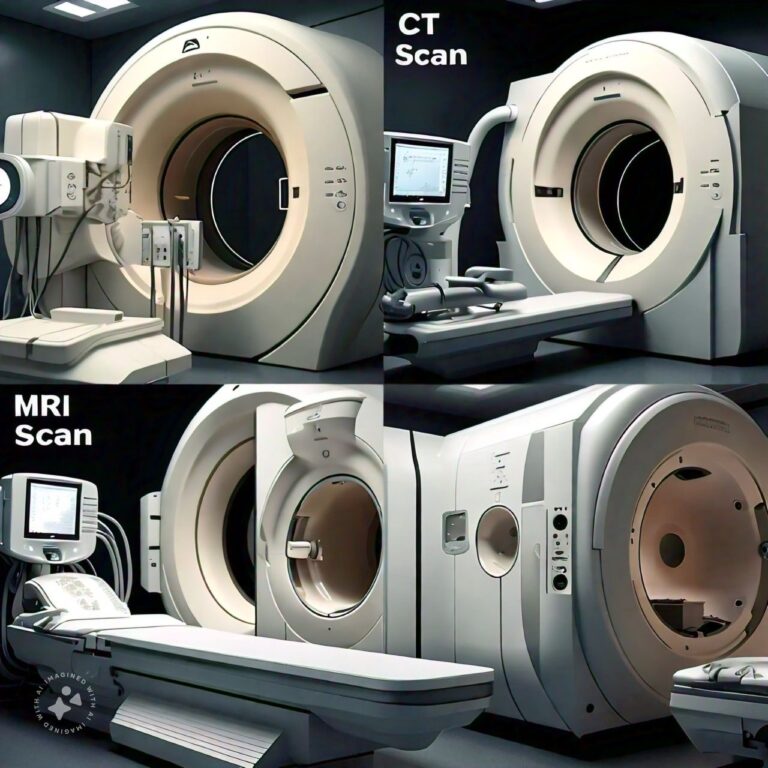
CT scans rely on X-rays, a form of ionizing radiation, to generate images. During the scan, the patient lies on a table that moves through a large, doughnut-shaped machine called a gantry. The gantry houses an X-ray tube that rotates around the patient, sending narrow beams of X-rays through the body from different angles. As the X-rays pass through the body, they are absorbed by different tissues in varying degrees.
Detectors on the opposite side of the X-ray tube measure the amount of X-rays that pass through the body and send this data to a computer. The computer processes the information to create detailed, cross-sectional images of the body. These images can be viewed as individual slices or reconstructed into 3D models to provide a more comprehensive view of the area being examined.
Types of CT Scans
There are several types of CT scans, each designed for specific purposes:
- Conventional CT Scan:
This is the standard CT scan, often used to examine different parts of the body, such as the head, chest, abdomen, and pelvis. - Helical (Spiral) CT Scan:
Helical CT scans involve continuous rotation of the X-ray tube, allowing for faster imaging and the ability to capture images of larger areas in less time. These scans are often used in trauma cases and cancer detection. - CT Angiography (CTA):
CT angiography uses contrast dye to visualize blood vessels and is commonly used to detect blockages, aneurysms, or other vascular abnormalities. - Low-Dose CT Scan:
This type of CT scan uses a lower dose of radiation and is often used for lung cancer screening or to assess patients at high risk of radiation exposure.
Benefits of CT Scans
CT scans offer several advantages, particularly in emergency settings and for the evaluation of bone and vascular conditions. Some key benefits include:
- Speed and Efficiency:
CT scans are quick, typically taking just a few minutes to complete, making them ideal for emergency situations like trauma or stroke. - Detailed Images:
CT scans provide highly detailed images of bones, blood vessels, and internal organs, making them useful for detecting fractures, tumors, and internal bleeding. - 3D Imaging:
CT scans can be reconstructed into 3D images, allowing doctors to visualize complex structures in more detail. - Non-invasive:
CT scans are non-invasive, meaning they do not require surgery or incisions.
Risks of CT Scans
Despite their usefulness, CT scans come with some risks, primarily due to the use of ionizing radiation. These risks include:
- Radiation Exposure:
CT scans expose patients to higher levels of radiation than standard X-rays, which can increase the risk of cancer over time, particularly with repeated scans. - Allergic Reactions to Contrast Dye:
Some CT scans use a contrast dye to enhance images, and in rare cases, patients may experience allergic reactions to the dye.
When Are CT Scans Used?
CT scans are commonly used in a variety of medical situations, including:
- Emergency Situations:
To quickly assess injuries from trauma, such as fractures, internal bleeding, or organ damage. - Cancer Detection:
To detect and monitor tumors and assess the effectiveness of cancer treatment. - Vascular Conditions:
To identify blockages, blood clots, aneurysms, and other vascular abnormalities. - Abdominal Pain or Infections:
To diagnose conditions such as appendicitis, kidney stones, or abdominal infections.
MRI Scan Overview
Introduction to MRI Scan
Magnetic Resonance Imaging (MRI) is a medical imaging technique that uses powerful magnets, radio waves, and a computer to create detailed images of the inside of the body. Unlike CT scans, MRI does not use ionizing radiation, making it a safer option for certain populations, such as pregnant women or children. MRI scans are particularly useful for examining soft tissues, including the brain, spinal cord, muscles, tendons, and ligaments.
MRI is commonly used to diagnose and monitor conditions affecting the:
- Brain and spinal cord (e.g., multiple sclerosis, brain tumors, stroke)
- Joints and musculoskeletal system (e.g., torn ligaments, herniated discs)
- Heart and blood vessels (e.g., heart disease, vascular abnormalities)
- Abdominal organs (e.g., liver, kidneys, spleen)
How MRI Scans Work
MRI scans use a combination of a magnetic field and radiofrequency pulses to create images of the body. During an MRI scan, the patient lies on a table that slides into a large tube-like machine, which houses a strong magnet. The magnet causes the protons (hydrogen atoms) in the body’s tissues to align in the direction of the magnetic field.
When the machine sends radiofrequency pulses into the body, these protons are temporarily knocked out of alignment. As the protons return to their original alignment, they release energy in the form of radio waves. The MRI machine detects these signals and sends them to a computer, which processes the data to create detailed images of the body’s internal structures.
Different tissues release different amounts of energy, allowing MRI to create high-contrast images that distinguish between soft tissues, bones, and fluids.
Types of MRI Scans
MRI scans can be tailored to specific diagnostic needs. Some of the most common types of MRI scans include:
- Functional MRI (fMRI):
This type of MRI measures brain activity by detecting changes in blood flow. It is often used in research and to assess brain function before surgery. - Magnetic Resonance Angiography (MRA):
MRA focuses on imaging blood vessels and is used to detect aneurysms, blockages, and other vascular conditions. - Cardiac MRI:
This type of MRI is used to assess the structure and function of the heart, as well as detect conditions such as heart disease and myocardial infarction. - Musculoskeletal MRI:
This scan is used to evaluate joints, muscles, tendons, and ligaments, making it useful for diagnosing sports injuries and musculoskeletal disorders.
Benefits of MRI Scans
MRI scans offer several advantages, especially when it comes to imaging soft tissues and organs. Some of the key benefits include:
- No Radiation Exposure:
MRI does not use ionizing radiation, making it safer for repeated use, especially in children and pregnant women. - High-Contrast Images of Soft Tissues:
MRI provides unparalleled detail of soft tissues, making it the preferred method for imaging the brain, spinal cord, and joints. - Versatility:
MRI can be used to examine various parts of the body, including the brain, spine, heart, and abdominal organs. - Non-invasive:
Like CT scans, MRI is a non-invasive diagnostic tool.
Risks and Limitations of MRI Scans
While MRI scans are generally safe, there are some limitations and risks to consider:
- Magnetic Field Risks:
The powerful magnetic field can interfere with metal implants, such as pacemakers, cochlear implants, or certain types of prosthetic joints, making MRI unsuitable for some patients. - Claustrophobia:
Some patients experience claustrophobia or anxiety while inside the MRI machine due to its enclosed design and loud noises during the scan. - Contrast Agent Risks:
In some cases, a contrast agent (gadolinium) may be used to enhance the images. Although rare, some individuals may have an allergic reaction to the contrast dye.
When Are MRI Scans Used?
MRI scans are often used when detailed images of soft tissues are needed. Common uses include:
- Neurological Conditions:
To diagnose brain tumors, multiple sclerosis, stroke, and other neurological conditions. - Spinal Injuries or Diseases:
To assess herniated discs, spinal cord injuries, or degenerative diseases. - Joint Injuries:
To detect torn ligaments, cartilage damage, or other musculoskeletal injuries. - Heart and Blood Vessels:
To evaluate heart structure and function, as well as detect vascular abnormalities.
Differences Between CT Scan and MRI Scan
- Technology:
- CT Scan: Uses X-rays and computer technology to produce images.
- MRI Scan: Uses magnetic fields and radio waves to generate images.
- Radiation:
- CT Scan: Involves ionizing radiation, which can pose a risk with repeated exposure.
- MRI Scan: Does not use radiation, making it safer for certain populations.
- Best For:
- CT Scan: Ideal for imaging bones, detecting fractures, and assessing conditions involving the chest, abdomen, and pelvis.
- MRI Scan: Superior for imaging soft tissues, such as the brain, spinal cord, muscles, and tendons.
- Speed:
- CT Scan: Generally faster, taking only a few minutes to complete.
- MRI Scan: Slower, with some scans taking 30 to 60 minutes or longer.
- Use in Emergencies:
- CT Scan: Preferred in emergency situations due to its speed and ability to detect injuries, such as internal bleeding or fractures.
- MRI Scan: Not typically used in emergencies, as it takes longer and may not be necessary for urgent assessments.
- Suitability for Metal Implants:
- CT Scan: Safe for patients with metal implants, pacemakers, or other medical devices.
- MRI Scan: Not suitable for patients with certain metal implants or devices, as the magnetic field can interfere with them.
- Detail of Images:
- CT Scan: Provides excellent images of bones and is useful for detecting tumors, internal bleeding, and other conditions.
- MRI Scan: Provides higher resolution images of soft tissues, making it better for detecting ligament tears, brain abnormalities, and spinal cord injuries.
- Contrast Agents:
- CT Scan: May use iodine-based contrast dye, which can cause allergic reactions in some patients.
- MRI Scan: May use gadolinium contrast, which has a lower risk of allergic reactions.
- Cost:
- CT Scan: Generally less expensive than MRI.
- MRI Scan: More expensive due to the advanced technology used.
- Noise Level:
- CT Scan: Quieter and less confining than MRI.
- MRI Scan: Loud and can be uncomfortable due to the confined space.
Conclusion
In conclusion, CT scans and MRI scans are powerful diagnostic tools that provide detailed images of the body’s internal structures, but they differ in how they achieve this. CT scans use X-rays and are typically faster, making them ideal for emergencies, especially for detecting bone injuries, internal bleeding, and conditions involving the chest and abdomen. MRI scans, however, use magnetic fields and radio waves, making them better suited for imaging soft tissues such as the brain, muscles, and ligaments without the risk of radiation exposure.
Choosing between a CT scan and an MRI scan depends on the specific medical condition, the part of the body being examined, and the patient’s individual needs, such as sensitivity to radiation or the presence of metal implants. While both imaging techniques have their advantages, MRI is often favored for its detailed images of soft tissues, while CT scans are more widely used for their speed and versatility in a variety of medical conditions.
Ultimately, the decision to use a CT or MRI scan should be made by a healthcare provider based on the diagnostic needs of the patient, ensuring the most accurate and effective treatment plan.
FAQs
Related Topics
- All
- Animals
- Diseases
- Health
- Money
- Politics
© 2024 OnYelp.com. All rights reserved. Terms and Conditions | Contact Us | About us


